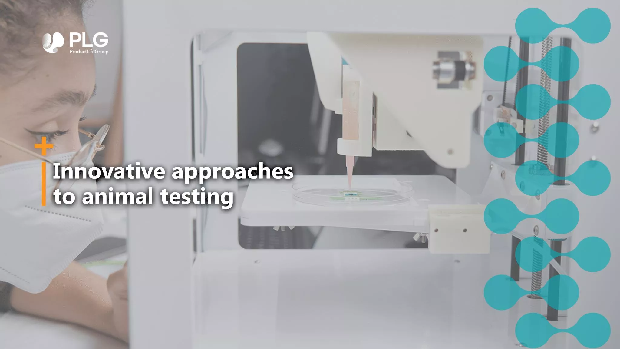
Innovative Approaches to Animal Testing
04 january 2024

Animal testing in its current form
The number of animals used in research has increased with the advancement of research and development in medical technology. Every year, millions of experimental animals are used all over the world. The pain, distress, and death experienced by the animals during scientific experiments have been a debated issue for a long time. Besides the major concern of ethics, there are a few more disadvantages of animal experimentation, like the requirement of skilled manpower, time-consuming protocols, and high cost. Various alternatives to animal testing were proposed to overcome the drawbacks associated with animal experiments and avoid unethical procedures. A strategy of 3 Rs (i.e., reduction, refinement, and replacement) is being applied for laboratory use of animals. Different methods and alternative organisms are applied to implement this strategy. These methods provide an alternative means for drug and chemical testing, up to some levels. An integrated application of these approaches would give an insight into the minimum use of animals in scientific experiments.
Three Rs: Reduction, Refinement and Replacement
- Reduction
With the help of statistical support and careful selection of study design, one can produce meaningful scientific results of an experiment. For example, in-vitro cell culture is a good way to screen the compounds at early stages. The human hepatocyte culture explains how a drug would be metabolized and eliminated from the body. Live animals and embryos are used to study the effects of some compounds on embryo development. In-vitro embryonic stem cell culture tests help reduce the number of live embryos and toxic compounds used to develop embryos.
- Refinement
Refining the animal facility reduces pain, discomfort, and distress during animal life and scientific procedures. Moreover, under stress and discomfort, there may be an imbalance in the hormonal levels of animals, leading to fluctuations in the results. Hence, experiments need to be repeated, which causes an increase in the number of experimental animals. So, refinement is necessary to improve laboratory animals’ lives and research quality.
- Replacement
Various alternatives to the use of animals have been suggested, such as in-vitro models, cell cultures, computer models, and new imaging/analyzing techniques. The in-vitro models allow studying the cellular response in a closed system, where the experimental conditions are maintained—for example, computer models, cell and tissue cultures, etc.
- Alternative methods to animal testing
Various methods have been suggested to avoid animal use in experimentation. These methods are described in detail as follows:
a) Computer models
Specialized computer models and software programs help to design new medicines. Computer-generated simulations predict the possible biological and toxic effects of a chemical or potential drug candidate without animal dissection. Only the most promising molecules obtained from primary screening are used for in-vivo experimentation.
Some such software models/ devices are included below:
- Computer Aided Drug Design (CADD)
The CADD method predicts the receptor binding site for a potential drug molecule. Another popular tool is the Structure Activity Relationship (SAR) computer program. It predicts the biological activity of a drug candidate based on the presence of chemical moieties attached to the parent compound. Quantitative Structure Activity Relationship (QSAR) is the mathematical description of the relationship between the physicochemical properties of a drug molecule and its biological activity. The recent QSAR software shows more appropriate results while predicting the carcinogenicity of any molecule.
- In-silico modeling
Another modeling method is in-silico modeling. In-silico modeling, in which computer models are developed to model a pharmacologic or physiologic process, is a logical extension of controlled in-vitro experimentation. In-silico modeling combines the advantages of both in-vivo and in-vitro experimentation without subjecting itself to ethical considerations and lack of control associated with in-vivo experiments.[1]
- 3D Bioprinting
3D bioprinting uses 3D printing–like techniques to combine cells, growth factors, and/or biomaterials to fabricate biomedical parts, often to imitate natural tissue characteristics. It has the potential to provide a unified framework for the manufacturing of tissue models for biomedical research, including drug discovery, disease modeling, and regenerative medicine.[2]
- Biochip
Biochip is an extensively studied and developed bio-microarray device to enable large-scale genomic, proteomic, and functional genomic analyses. A biochip comprises three types: DNA microarray, protein microarray, and microfluidic chip. With the integration of microarray and microfluidic systems, a micro total analysis system, often called a lab-on-a-chip (LOC) system, is produced. Due to the benefits of low expense, high throughput, and miniaturization, this technology has great potential to be a crucial and powerful tool for clinical research, diagnostics, drug development, toxicology studies, and patient selection for clinical trials.[3]
b) Cells and tissue cultures
In-vitro culture of animal/human cells includes their isolation from each other and growing as a monolayer over the surface of culture plates/flasks. Cellular components like membrane fragments and cellular enzymes can also be used. Various types of cultures like cell, callus, tissue, and organ cultures are used for various purposes.
2D cell culture and pre-clinical animal models have traditionally been implemented to investigate the underlying cellular mechanisms of human disease progression. However, the increasing significance of 3D versus 2D cell culture has initiated a new era in cell culture research in which 3D in-vitro models are emerging as a bridge between traditional 2D cell culture and in-vivo animal models. Additive manufacturing (AM, also known as 3D printing), defined as the layer-by-layer fabrication of parts directed by digital information from a 3D computer-aided design (CAD) file, offers the advantages of simultaneous rapid prototyping and biofunctionalization as well as the precise placement of cells and extracellular matrix with high resolution.[4]
Additionally, organoids and spheroids are different 3D cell culture models that can be cultured with different techniques. These 3D cell culture units established from a patient tumor have similarities to the original tumor tissue and possess several advantages in conducting basic and clinical cancer research.[5]
Recently, organs-on-chips (OoCs) have been found to contain engineered or natural miniature tissues grown inside microfluidic chips. To better mimic human physiology, the chips are designed to control cell microenvironments and maintain tissue-specific functions. Combining advances in tissue engineering and microfabrication, OoCs have gained interest as a next-generation experimental platform to investigate human pathophysiology and the effect of therapeutics in the body.[6]
c) Alternative organisms
Some alternative organisms that can be used are listed below:
- Lower vertebrates
Lower vertebrates are an attractive option because of their genetic relatedness to the higher vertebrates, including mammals. (e.g., zebrafish).
- Invertebrates
Various diseases like Parkinson’s disease, endocrine and memory dysfunction, muscle dystrophy, wound healing, cell aging, programmed cell death, retrovirus biology, diabetes, and toxicological testing. They hold numerous benefits, such as a brief life cycle, small size, and simple anatomy, so that many invertebrates can be studied in a single experiment within a short period with fewer ethical problems and exampled by Drosophila melanogaster (fruit fly), Caenorhabditis elegans (eukaryotic nematode).
- Microorganisms
For example, Saccharomyces cerevisiae (Brewing yeast) is the most popular and important model organism due to its rapid growth, ease of replica plating and mutant isolation, dispersed cells, well-defined genetic system, and highly versatile DNA transformation system.
The future of animal testing in pre-clinical phase
Replacing animal tests does not mean putting human patients at risk. It also does not mean halting medical progress. Instead, replacing animals used in testing will improve the quality and humanity of our science. Thankfully, the development of non-animal methods is growing fast. Due to innovations in science, animal tests are being replaced in areas such as toxicity testing, neuroscience, and drug development. But much more needs to be done.
Bioinformatics tools, in-vitro cell cultures, enzymatic screens, and model organisms are necessary to integrate various computer models. Using modern analytical techniques, data acquisition, and statistical procedures to analyze the results of alternative protocols can provide dependable outcomes. These integrated approaches would result in minimum involvement of animals in scientific procedures.
Product life group can provide valuable support in strategizing appropriate alternative methods for your planned animal testing, considering the regulatory possibilities.
References:
- Colquitt RB, Colquhoun DA, Thiele RH. In silico modelling of physiologic systems. Best practice & research Clinical anaesthesiology. 2011 Dec 1;25(4):499-510.
- Pless CJ, Radeke C, Cakal SD, Kajtez J, Pasqualini FS, Lind JU. Emerging strategies in 3D printed tissue models for in vitro biomedical research. InBioprinting 2022 Jan 1 (pp. 207-246). Academic Press.
- Zhang X, Ju H, Wang J, editors. Electrochemical sensors, biosensors and their biomedical applications. Academic Press; 2011 Apr 28.
- Vanderburgh J, Sterling JA, Guelcher SA. 3D printing of tissue engineered constructs for in vitro modeling of disease progression and drug screening. Annals of biomedical engineering. 2017 Jan;45:164-79.
- Gunti S, Hoke AT, Vu KP, London Jr NR. Organoid and spheroid tumor models: Techniques and applications. Cancers. 2021 Feb 19;13(4):874.
- Leung CM, De Haan P, Ronaldson-Bouchard K, Kim GA, Ko J, Rho HS, Chen Z, Habibovic P, Jeon NL, Takayama S, Shuler ML. A guide to the organ-on-a-chip. Nature Reviews Methods Primers. 2022 May 12;2(1):33.
Register to our news and events
Go to our Events to register
Go to our News to get insights
